


Chest pain
- Chest pain: definition
- The steps diagnosis of chest pain
- The main examinations
- The differential diagnosis of chest pain
- Conclusion
1. Chest pain: definition
Chest pain is the symptom which representes:
approximately 30% of subjective troubles motivating medical consultation
more than 50% of reasons for hospitalizations in cardiology.
Its etiology is extremely varied and dominated by cardiovascular causes, including three critical emergencies that must be quickly eliminated:
myocardial infarction
pulmonary embolism
aortic dissection
Depending on the location of chest pain, we talk about:
Retrosternal pain: behind the sternum, eg in the middle of the chest
Latero thoracic pain: on the side of the thorax.
Basi thoracic pain: at the base of the thorax.
A detailed examination and a thorough clinical examination of the patient can usually clarify the diagnosis
2. The steps diagnosis of chest pain
The interview and physical examination of the patient
It will specify:
- the type of pain (constrictive, burning, crushing, pinching, cutting with knives)
- the mode of onset pain (spontaneous, effort or equivalent postural)
- duration, frequency and intensity of pain (start time, any paroxysms)
- the seniority pain and the pain development (acute or chronic pain)
- the proneness to taking drugs (cédocard)
- the pain location (retrosternal, basithoracique)
- the pain irradiation (upper limbs, jaw, back)
- any pain reproduction on palpation or on movement of the patient
- the clinical signs accompanying pain (dyspnea, palpitations, cyanosis, faintness, fever, digestive disorders)
- the attitudes that relieve pain (position leaning forward, ...)
The search for clinical signs of severity
- Difficulty breathing
- Associated symptoms: digestive intolerance, dizziness, syncope
- Disturbance of consciousness
- Weak peripheral pulse (shock)
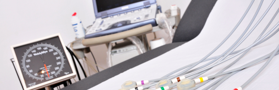
3. The main examinations
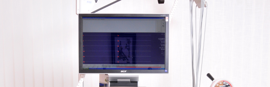
- Electrocardiogram: repeat if necessary
- Chest X-ray
- Cardiac Ultrasound
- Biological analysis: determination of troponin or high sensitive troponin (HS troponin)
- Other tests requested, according to the clinical finding
4. The differential diagnosis of chest pain
Many organic or functional disorders can cause chest pain:
- Chest pain of cardiovascular origin
- The coronary artery disease
- Pulmonary embolism
- Aortic dissection
- Acute pericarditis
- Chest pain of non-cardiovascular origin
- The pain of origin pulmonary (pneumothorax, pleurisy)
- The pain of gastro-esophageal origin
- The pain of abdominal origin
- The Parietal pain of rheumatic origin
- The pain of neurological origin
- False anginal pain of psychogenic origin
- Other causes of chest pain
There is no formal parallelism between the severity of the affection and the pain intensity felt by the patient!
Chest pain of cardiovascular origin
Four causes of chest pain to systematically search because of their immediate or potential severity:
- coronary artery disease
- pulmonary embolism
- aortic dissection (tear in the wall of the aorta)
- Acute pericarditis
Any prolonged chest pain, occurring in a patient older than 40 years with risk factors of cardiovascular disease requires the execution of an electrocardiogram (ECG) and hospitalization for clinical monitoring.
The coronary artery disease
Coronary artery origin should be suspected with the association of three factors:
- typical chest pain : constrictive, mid-chest, radiating to the arm or jaw...
- evocative area : coronary personal history, family, diabetes, smoking, high cholesterol, hypertension
- suggestive ECG signs: repolarization disorders, infarction sequelae
In case of ECG signs compatible with acute coronary insufficiency, hospitalization is justified for urgent initiation of treatment and the patient monitoring.
A normal ECG and biomarkers of myocardial infarction during the first normal dosage do not eliminate the diagnosis of coronary artery disease, particularly if there are risk factors and if the chest pain is typical. In any doubt, a biological testing should be performed to determine the biological markers of myocardial infarction (troponin, CPK).
Pulmonary embolism
The diagnosis of pulmonary embolism should be suspected when a chest pain, associated with sudden dyspnea (difficulty breathing) occurs in a patient who has a phlebitis or risk factors (post-partum, post-operative, association and pill smoking, heart failure ...).
Chest pain can be sided, but it may be in the middle of the chest, mimicking an acute coronary insufficiency.
Paraclinical examinations:
- ECG: The ECG sign is the most common sinus tachycardia. Other signs are more suggestive ECG (S1 Q3, right bundle branch block, negative T wave in the right derivations).
- Cardiac Ultrasound: It is used to evaluate the severity of the disease by investigating the impact on the right heart (pulmonary hypertension, enlargement of right cavities)
- Biological analysis: determination of D-dimers. If the dosage of D-dimer is negative, the diagnosis is ruled out with a very good negative predictive value (97%). A positive test is not very specific (66%) for the presence of a pulmonary embolism..
- The blood gas analysis or gas: It must be conducted to assess the severity of pulmonary embolism. It shows a decrease in CO2 pressure (hypocapnia) and hypoxemia (reduced oxygen pressure).
- The scintigraphy of lung ventilation and perfusion or the CT scan angio-spiral: They can be conducted to confirm the diagnosis and assess the severity of the condition.
- The chest X-ray: It may show atelectasis in strips or pleural basal effusion.
In case of suspicion of the pulmonary embolism diagnosis, treatment with effective dose of heparin, must be started before confirmation of the diagnosis.
Aortic dissection
Aortic dissection should be suspected with a sharp pain in the beginning, felt like a stab, prolonged and radiating in the back.
Some specific clinical signs should be sought
- emergence of a aortic breath
- asymmetry of blood pressure in both arms
- shock
- neurological deficit, ...
The factors:
- A former high blood pressure poorly controlled
- Marfan syndrome
- thoracic trauma
Paraclinical examinations:
- The ECG : is usually normal, thus eliminating the diagnosis of myocardial infarction.
- The chest X-ray: An enlargement of the upper mediastinum is observed.
In case of doubt about the diagnosis of aortic dissection, cardiac ultrasound trans-oesophageal must be made, possibly supplemented by a scanner or an MRI chest.
Acute pericarditis
The diagnosis of acute pericarditis is referred to a chest pain with certain clinical characteristics:
- The pain is modified by respiration and changing positions
- A viral context is associated or has proceeded the onset of symptoms
- Clinical examination revealed the presence of fever or a pericardial friction fleeting.
Paraclinical examinations:
- The ECG: It shows an elevation of ST segment, concave and diffuse.
- Biological analysis: It shows the presence of an inflammatory syndrome. Biomarkers of myocardial infarction may be moderately high in cases of inflamed heart muscle (myo-pericarditis).
- The chest X-ray: It sometimes shows an enlargement of the cardiac silhouette.
- Cardiac Ultrasound: In the absence of the typical table (pain + fever + friction), a cardiac ultrasound may confirm the diagnosis by demonstrating a pericardial effusion. In this case, the clinical setting can often make the difference between pericarditis and myocardial infarction in evolution.
Pericarditis requires hospitalization for:
- cardiac assessment for etiology
- the initiation of treatment
- the patient clinical monitoring (risk of rare but serious tamponade)
Chest pain from non-cardiovascular
The pain of origin pulmonary (pneumothorax, pleurisy)
Pain:
- sided
- sudden onset
- increases with breathing and coughing.
- associated with dyspnea or a polypnea
There may be an underlying infection (fever, chills)
Paraclinical examinations:
- The chest radiograph: To clarify the diagnosis (pulmonary condensation, presence of air in the pleural space)
The pain of gastro-esophageal origin
It is felt as a bottom-burning, relieved by the dressing gastric (heartburn).
Esophageal spasm can be felt as a pseudo-anginal pain triggered by swallowing, of short duration, relieved by taking nitrates (cédocard).
Paraclinical examinations:
- The esophageal manometry: Shows esophageal dyskinesia.
The intricacy of the clinical symptoms of esophageal spasm with angina is an authentic as possible.
The different causes of pain of gastro-esophageal origin:
- gastric ulcer
- rupture of the esophagus
- for gastroesophageal reflux disease
- achalasia
A detailed analysis of patient Complaints is fundamental and requires a specialized cardiological opinion if doubt!
The pain of abdominal origin
They are caused mainly by:
- the gallbladder stones
- acute pancreatitis
- peritonitis
- the presence of a sub-phrenic abscess
Colic liver can cause chest pain basi-straight, sometimes with radiation to the right shoulder.
Some diseases of the spleen are pain basi-left chest, can radiate to the left shoulder.
The origin of pancreatic pain is typically epigastric, transfixiante and sometimes described as retrosternal.
The Parietal pain of rheumatic origin
- Tietze's syndrome : mechanical pain of articulation sterno-costal with local swelling
- (ligament) Musculoskeletal pain: insertion of the major pectoralis muscle on the sternum
- Sternal lesions (infectious, neoplastic)
The pain of neurological origin
They root in belt, exacerbated by coughing.
They can be caused by:
- settling vertebral
- malignant tumors
- the neuroma,
- intercostal shingles,
- Lyme disease (radiculitis)
False anginal pain of psychogenic origin
They are extremely common and form the basis of extensive consultations.
These pains:
- can be located with precision on the thorax,
- occur pin coup or stabbing coup,
- are often left latero-thoracic or straight or localized in the breast region.
- may be extremely brief (a few seconds to several minutes), sometimes accented by a deep inspiration,
- may be precipitated by a movement of the trunk or arms.
The accompanying signs such as anxiety, neurosis or palpitations, are frequently associated. Their duration is relatively short. They generally have no particular trigger
Other causes of chest pain
- Mediastinal: mediastinal tumors, pneumomédiastin, mediastinitis
- Chest pain as part of a rhythm disorder
- Rupture of an aneurysm in the sinus of Valsalva
Chest pain is an important symptom that may be indicative of a serious illness, requiring treatment in emergency (myocardial infarction, pulmonary embolism and aortic dissection).
The causes of chest pain are extremely varied. A medical consultation is often necessary to clarify the diagnosis and require a thorough examination, a clinical examination or tests.
Any suspicious chest pain and prolonged (more than ten minutes), occurring in patients with cardiovascular risk factors requires a medical emergency.